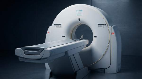
X-ray computed tomography (CT) lets researchers see inside an object in three dimensions without cutting it open. That single property—non-destructive 3D imaging—distinguishes it from microscopy techniques that require sectioning, such as scanning electron microscopy or transmission electron microscopy, and it is the reason the method has spread from hospital radiology into laboratory benches studying welds, batteries, and rodent hearts. The trade-off is that CT is not equally good at everything: laboratory micro-CT prioritizes resolution over field of view and speed, dense materials image more readily than soft tissue, and biological samples carry a radiation-dose budget that must be managed. Understanding the physics and the reconstruction pipeline explains why those trade-offs exist.
Understanding X-ray Computed Tomography: Core Principles
Every CT scan begins with an X-ray source. X-rays are generated and directed through the sample, where they are attenuated—that is, absorbed or scattered—to different degrees depending on the density and composition of the material they encounter. Denser regions such as bone, metal, or mineralized tissue attenuate more X-rays than voids, pores, or soft tissue. A detector on the far side of the sample measures the X-rays that survive, converting the transmitted signal into digital data. A single pass through the sample, however, produces only a two-dimensional shadow: overlapping structures are superimposed and cannot be separated.
The solution is rotation and computation. The object is rotated between the source and detector—or, in some geometries, the source and detector rotate around the object—and many projection images are acquired from hundreds of angles. Each projection records a different set of attenuation path-lengths through the sample. Reconstruction algorithms then solve the inverse problem of recovering the internal attenuation distribution from this stack of projections, a mathematical relationship formalized in the Radon transform, which underpins CT reconstruction as described in a review of CT's basic physical principles in Progress in Medical Physics. The output is a stack of virtual cross-sectional "slices" that can be stacked into a full 3D volume.
The result is what makes CT distinctive: you can scan an object and get slices that reveal internal structures without physically slicing the object, as instrument maker Rigaku puts it in its technical introduction, noting that this 3D capability without sectioning is the technique's biggest advantage over other microscopy methods. The same review of CT principles in a 2026 advanced-materials survey in the Journal of South China University of Technology identifies the core pipeline as X-ray attenuation, signal conversion, and 3D reconstruction. Everything that follows—defect detection, porosity measurement, anatomical imaging—is an application of that pipeline with different parameters.
X-ray CT in Materials Science: Analyzing Internal Structures
In materials science, CT answers questions about hidden internal geometry. The advanced-materials survey documents case studies in weld defect detection in aerospace components, hidden defect identification in electronic packaging solder joints, and porosity quantification in additive manufacturing materials. In each case the goal is the same class of problem: locate and measure features that are invisible from the surface and would be destroyed or altered by conventional cross-sectioning. CT provides defect localization, damage tracking, and microstructure quantification as quantitative 3D datasets rather than single-plane micrographs.
The technique also supports dynamic and electrochemical studies. The same survey highlights applications in lithium-ion battery electrode evolution, fuel cell water management, and metal anode dendrite tracking—experiments where the value lies in watching internal structure change over time, revealing the relationship between material structure and electrochemical performance, informing device design, and improving safety. Repeated scanning of the same specimen is only possible because the measurement is non-destructive.
Resolution is where laboratory CT separates itself from clinical scanners. Medical CT resolves at submillimeter to millimeter scale, whereas micro-CT and nano-CT scanners achieve micrometer and sub-micron resolution respectively, with nano-CT sometimes described as X-ray microscopy because its resolution approaches that of optical microscopes. Common micro-CT scanners can image objects up to roughly 20 cm in diameter with pixel sizes in the micrometer range. That resolution advantage comes at the cost of smaller fields of view and slower acquisition, a design trade-off rather than a defect.
X-ray CT in Biological Research: Non-destructive Sample Analysis
Biological applications split into two regimes. For ex vivo work—isolated, fixed specimens—micro-CT delivers exceptional detail. A Duke University dissertation on micro-CT principles reports imaging a fixed rodent heart at 25 µm spatial resolution, producing a 3D reconstruction with superb anatomical detail. Because microfocus X-ray sources require longer exposure times to reach adequate photon fluence and signal-to-noise ratio, this high resolution is most practical when radiation dose is not a concern and when the specimen is motionless, which fixed samples are by definition.
For in vivo work, micro-CT extends to small animal imaging—mice and rats, and in some cases rabbits—again without sectioning, preserving specimens for future studies. But living samples introduce two complications that fixed specimens do not have. First, motion: samples must remain motionless during acquisition to avoid blurring, so in vivo imaging is best suited to specimens with minimal biological motion. Second, dose: repeated scanning of living animals accumulates radiation that could affect biological function, particularly immune function and tumor response, and long-term health.
Contrast is the third biological consideration. Because the X-ray signal is density dependent, micro-CT readily visualizes mineralized or dense features such as bone, but soft tissue is another matter. Recent advances in improving soft-tissue contrast through heavy-metal staining, as summarized in ScienceDirect's topic overview, are expanding the technique's applicability, and for polymer and biological samples the dose cannot exceed a limit because extended scans can destroy samples through radiation damage.
Comparative Applications of X-ray CT in Materials Science vs. Biological Research
The table below contrasts the two domains across four dimensions: what CT is used for, what scientific question it answers, what resolution it typically delivers, and the defining advantage or limitation in that context. The comparison shows how one physical technique is re-parameterized for very different problems—and where each domain hits its own boundary.
| Dimension | Materials Science | Biological Research |
|---|---|---|
| Application Area | Defect localization, damage tracking, and microstructure quantification in welds of aerospace components, electronic packaging solder joints, and additive manufacturing materials. | Small animal imaging (mice, rats, and in some cases rabbits) and imaging of isolated, fixed specimens such as rodent hearts, without sectioning. |
| Scientific Goal | Reveal relationships between material structure and performance—e.g., lithium-ion battery electrode evolution, fuel cell water management, and metal anode dendrite tracking—to optimize design and improve safety. | Visualize mineralized or dense features; soft-tissue contrast is harder and may require heavy-metal staining, with protocols designed to minimize radiation effects. |
| Typical Resolution | Micrometer-range pixel sizes for micro-CT, sub-micron for nano-CT—far beyond the submillimeter-to-millimeter resolution of medical CT. | High-resolution micro-CT, achieving spatial resolutions such as 25 µm for ex vivo rodent heart specimens, yielding detailed 3D anatomy. |
| Key Advantage/Limitation | Non-destructive, 3D quantitative, and capable of dynamic tracking; limited by low efficiency in nanoscale imaging and difficulties in data fusion. | Non-destructive analysis preserves samples for future studies; limited by cumulative radiation dose effects on biological function and motion artifacts in living specimens. |
Read across the rows, the pattern is consistent: materials science exploits CT's tolerance for dense samples and high dose to push resolution and dynamic tracking, accepting nanoscale inefficiency and data-fusion difficulty as bottlenecks. Biology exploits the same non-destructiveness to preserve irreplaceable specimens, but must budget dose, control motion, and often stain for soft-tissue contrast. Neither domain gets everything; each optimizes the parameters that matter most for its samples.
Key Limitations and Practical Considerations for X-ray CT
Sample size and density set the first boundary. Micro-CT's micrometer resolution comes with a bore-size and field-of-view penalty—common scanners handle objects up to about 20 cm in diameter—and the density-dependent signal means highly attenuating metals and mineralized tissue image readily while polymers and soft tissue yield weak absorption contrast. Where staining with heavy elements is possible, contrast improves; where it is not, absorption-contrast imaging of polymers at high spatial resolution becomes unreliable.
Resolution has its own ceiling. Nano-CT reaches sub-micron resolution, but the advanced-materials survey identifies low efficiency in nanoscale imaging as a bottleneck, and combining CT data with other measurement modalities raises data-fusion difficulties. Higher resolution also demands more photons: microfocus sources need longer exposure times to achieve the fluence and signal-to-noise ratio that support fine low-contrast detail, which directly increases scan time.
For biological samples, dose is the binding constraint. The Duke micro-CT review stresses that careful consideration must determine the optimal imaging protocol for each individual application to minimize radiation-dose effects on the experiment, and that polymer and biological samples can be destroyed by radiation damage during extended scans. The review also notes that advances in micro-CT technology and reconstruction algorithms should further reduce doses, gradually eroding this limitation. Motion completes the picture: any movement during acquisition blurs the reconstruction, so accurate resolution requires motionless objects—trivially true for fixed specimens, actively managed in living ones.
Deciding on X-ray CT for Your Research
Researchers should evaluate X-ray CT's suitability by considering their specific scientific goal, the nature of their sample (size, density, biological sensitivity), and the required spatial resolution, while acknowledging limitations like radiation dose and potential motion artifacts. A dense material with a defect-detection question and a micrometer-scale target is squarely in CT's strength; a soft, radiation-sensitive, moving biological target demands staining, dose budgeting, and protocol design before committing. The measurable indicator of a good fit is the successful acquisition of high-resolution 3D internal structural data that directly addresses the research question without compromising sample integrity or introducing significant artifacts. If a planned protocol cannot plausibly deliver that—because the dose would damage the specimen, the motion would blur the slices, or the required resolution exceeds what micro-CT efficiently delivers—then the technique, or the protocol, needs rethinking before the first scan.
Sources
- Basic Physical Principles and Clinical Applications of Computed Tomography — Progress in Medical Physic
- X-ray Computed Tomography: A Brief Introduction — RIGAKU
- Application of X-Ray Computed Tomography in Frontier Research of Advanced Materials — SciOpen
- Laboratory x-ray micro-computed tomography: a user guideline for biological samples - PMC
- Dukespace
- Sciencedirect